Growing role in diagnosis and monitoring of infant and young child eye conditions. Howard Larkin reports
Howard Larkin
Published: Wednesday, September 1, 2021
Hand-held OCT (HH-OCT) provides a unique in vivo view of the eye’s early development, enabling rapid diagnosis of paediatric ocular conditions ranging from differential aetiology of nystagmus to early-onset retinal dystrophies and optic nerve diseases such as glioma, glaucoma, and optic nerve hypoplasia, Irene Gottlob MD, FRCOphth said in a presentation reviewing work by her group and others at the Association for Research in Vision and Ophthalmology 2021 Annual Meeting.
HH-OCT allows the technology to be used effectively on infants and young children by adjusting the hand-held device to the child’s position.
“Very small children, even premature children, can be examined,” said Dr Gottlob, University of Leicester, UK.
This enables viewing in real-time the development of the retina only previously known through sporadic histology samples. For example, the growing differentiation of retinal pigment epithelium and photoreceptor cells, and development of the foveal pit as well as thickening of the outer nuclear layer under it, can readily be tracked as the retina matures, Dr Gottlob noted. Development continues until at least age 12, if not adulthood, and closely parallels development of the visual cortex and visual acuity, she added.
DIFFERENTIAL DIAGNOSIS IN NYSTAGMUS
Nystagmus is associated with foveal hypoplasia often clearly visible on OCT, Dr Gottlob said. Albinism and aniridia display similar “typical” foveal hypoplasia patterns, with the foveal pit shallow or missing. Achromatopsia can be distinguished on OCT by its “atypical” foveal hypoplasia, including pathological continuation of the inner retinal layers, disrupted inner segment/outer segment junction, and “punched out” hyporeflective zones where central photoreceptors should be that grow with age.
Retinal dystrophies can be identified by abnormal lamination of the outer retinal layers, Dr Gottlob said. Retinal pigment epithelium (RPE) may not be distinguishable from photoreceptors compared with normal control images.
A grading system for foveal hypoplasia based on OCT images shows increasing severity corresponding to reduced visual acuity, Dr Gottlob said. This system was able to predict future VA, demonstrating the technology’s diagnostic and prognostic utility.
RETINAL AND OPTIC NERVE IMAGING
In retinoblastoma, OCT can be used to increase diagnostic accuracy and clinical staging, resulting in changes in management in 15% of cases in one study, Dr Gottlob said. Retinal or optic nerve abnormalities are also visible in the following cases: microcephaly; optic nerve drusen; and optic nerve hypoplasia. Optic nerve changes can be monitored in optic nerve gliomas and childhood glaucoma. In glaucoma, corneal changes and iridocorneal angle changes can also be observed.
Optic nerve head pits and related retinal disruptions are also easily seen on OCT, Dr Gottlob said. She also pointed out that optic nerve abnormalities are visible in children with congenital fibrosis of the extraocular muscles, which confirms this disease affects wider parts of the eye and not only the extraocular muscles.
Irene Gottlob: ig15@leicester.ac.uk
Image 1: The hand-held OCT probe is pointed to the child’s eye, enabling scans in children who

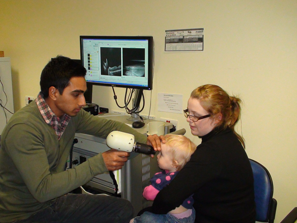 Image 1: The hand-held OCT probe is pointed to the child’s eye, enabling scans in children who